FOOT AND ANKLE PAIN SPECIALISTS SERVING MARLTON, N.J. AND RIDLEY PARK, PA.
TYPES OF FOOT AND ANKLE PAIN
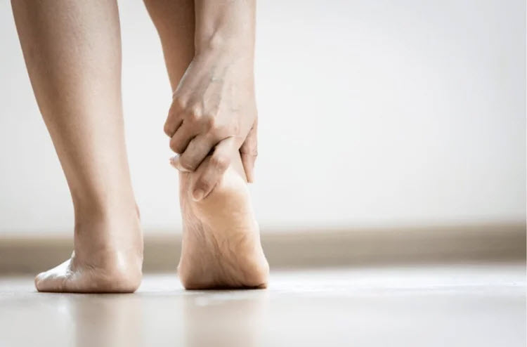
HEEL PAIN - PLANTAR FASCIITIS

The most common heel pain seen in our office is known as plantar fasciitis which can occur in athletes as well as the general population. The plantar fascia is a three-band thick ligament-like tissue that supports the bottom (or plantar surface) of the foot. It starts in the heel and spans the length of the foot to the toes.
Plantar Fasciitis is an inflammatory response due to exercise pressure in the arch and heel area caused by poor foot mechanics caused by improper walking or running. This inflammatory process usually comes on insidiously without warning. Highly active people such as runners can develop the problem after time, but it can occur in anyone, both male or female. There is some correlation with the frequency of plantar fasciitis in people who are overweight or obese.
How is Plantar Fasciitis Diagnosed?
Plantar fasciitis is pretty simple to diagnose with a physical exam. We look at your foot type, mechanics and we evaluate your heel and arch for painful areas. Sometimes in an x-ray, we see a bone spur on the heel. Usually, you have most of your pain in your first step out of bed in the am or after sitting for a while then returning to activity.
If the physical exam is not enough to diagnose the cause of your heel pain, your doctor may order x-rays of your foot to confirm there is no stress fracture or other issue that could be causing the pain.
Plantar Fasciitis Treatment
We approach each patient a little bit differently according to the results of their examination. Stretching, anti-inflammatory manipulation therapy such as active release techniques (ART), a form of deep tissue massage, as well as injection therapy for severe or more painful cases.
The main treatment objective is to realign the body and posture so that the foot functions properly without stressing the plantar fasciitis. This is accomplished through custom-made orthotic insoles. Our insoles are unique in that we have eight different corrective supportive cushion density materials with which we construct our custom insoles. Most other custom orthotics provide only one type of material.: Custom orthotics or heel pads may be utilized to help cushion the foot and prevent pain.
More severe cases of plantar fasciitis might require a surgical lengthening of the plantar fascia to relieve the pressure in addition to the custom orthotic insole. We have found a very helpful non-invasive technique that we do under anesthesia called shockwave therapy. Surgery is often seen as a last resort after conservative approaches have failed.
Contact Dr. Lee S. Cohen & Associates today, and we'll get you back, to the activities you love, in no time.
COMMON SYMPTOMS, CAUSES, AND TREATMENT OF FOOT ARCH AND HEEL PAIN
FOOT ARCH PAIN

What is a Foot Arch?
Foot arches, located in the area between the heel and ball of the foot, are shaped by bones and supported by tendons and ligaments (which stabilize the joints and help to keep the bones in place). There are two arches in your foot that crisscross one another: The longitudinal arch which runs up and down the foot and the transverse arch which extends across the width of the foot.
Arch Pain Symptoms
Arch pain can feel like a tightness, pulling, or a burning sensation on the bottom of your foot, typically in the ball of the foot and heel areas. Other symptoms can include swelling, stiffness, redness, and inflammation which can also coincide with the above.
Understanding Arch Pain
Arch pain may be felt in one or both arches and can be caused by one or several conditions that develop in the sole of your foot. However, it is important to recognize that you may be feeling related pain elsewhere, along the kinetic chain, since the proper functioning of your ankles, knees, hips, and back depends on a good stable foot foundation.
Arch Pain Causes
Injuries, overuse, genetics, or faulty biomechanics are some of the most common causes of arch pain, specifically:
- Flatfeet
- High-arched Feet
- Plantar Fasciitis
- Tendonitis
- Sprains and Strains
- Stress Fractures
- Pronation
- Overuse
Certain Types of Arthritis
Lack of stretching, improper exercise, and improper footwear selection are also some common contributing factors.
Arch Pain Diagnosis and Treatment
Determining which part of the foot is affected and what is causing the pain is the first step to treating arch pain. To do this, one of our sports medicine specialists will perform a full biomechanical exam and possibly a series of X-rays or 3-d imaging scans, allowing them to pinpoint and diagnose the issue.
Common treatment solutions may include a combination of one or more of the following:
- Anti-Inflammatory Medication
- Stretching
- Footwear Modifications
- Custom Orthotics
If you or someone you know is experiencing arch pain, foot pain, or pain in the areas of the lower body extremities (foot, ankle, knee, leg, hip, or back), call us to schedule an examination with one of our sports medicine podiatrists today calling 610-522-9200 or by using our contact form.
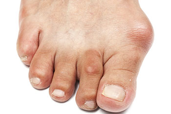
HEEL SPUR
What is a Heel Spur?
A heel spur is a calcium deposit found under the heel that causes small pieces of bone to protrude. Pain associated with heel spurs can often be confused with another condition called plantar fasciitis—which refers to inflammation in the plantar fascia ligament. A heel spur, on the other hand, is a piece of bone that forms on the heel bone itself.
In most cases, heel spurs do not directly cause any symptoms. Although there are some cases where heel spurs are associated with intermittent or chronic pain. However, the heel spur itself is not necessarily the root of the pain. Instead, the pain is attributed to the inflammation or irritation of the plantar fascia ligament. Pain is usually worse in the morning when you first wake up, but it dissipates as ligaments loosen.
Heel Spur Causes
Heel spurs can take months to form and may go completely undetected. Heel spurs are typically a result of too much stress or pressure on the ligaments. They can also be the result of continuous tearing of the membrane that covers the heel bone. The same physical activities that cause plantar fasciitis can also cause heel spurs.
Other contributing factors in the development of heel spurs include:
- Mechanical deficiencies that cause gait abnormalities
- Tight calf muscles that limit ankle mobility
- Obesity or being overweight
- Poorly chosen footwear
- Extended time spent on your feet
Treatment Options
Heel spurs are not easily diagnosed through a physical exam, because they can only be detected using an x-ray. Many heel spurs are found in the images your doctor takes while looking for something else. When heel pain continues for more than a month, you need to consult your podiatrist. He or she may recommend the following non-invasive treatment solutions:
- A variety of stretching exercises
- Shoe replacement
- Custom orthotics
- Physical therapy
Most cases of heel pain can be treated conservatively, but anti-inflammatory medications or injections may also be considered as a treatment option. These medications not only help to mitigate pain but help to reduce inflammation as well. Contact Dr. Lee S. Cohen & Associates today for a complete evaluation.
HAGLUND’S DEFORMITY

What is Haglund’s Deformity?
Also known as the “pump bump”, Haglund’s deformity is a condition that causes the bone section of your heel—where the Achilles tendon is—to become enlarged or inflamed. Wearing shoes that put too much pressure on the back of the heel, which causes inflammation, most often causes Haglund’s deformity.
If left untreated, Haglund’s deformity can lead to bursitis—which is an inflammation of the fluid-filled sac that separates the tendon from the bone. When the heel becomes inflamed, it can actually calcify the heel bone and cause the bump to become more prominent. When this happens, pain becomes more noticeable and basic foot function could be affected.
Causes of Haglund’s Deformity
As its other name “pump bump” implies, the rigid backs of “pump-style” shoes usually cause Haglund’s deformity. These shoes create pressure that aggravates the growth during normal activities, like walking.
In addition to poor shoe choice, these factors can also contribute to the formation of Haglund’s deformity:
- Having a high-arched foot
- Having a tight Achilles tendon
- Poor walking mechanics
Haglund’s Deformity Symptoms
Most cases of Haglund’s deformity are very painful—especially in the area where the growth is located o the heel. Other common symptoms of Haglund’s deformity include:
- Noticeable bump on the back of the heel
- Severe pain in the heel
- Swelling in the heel
- Redness or tenderness near the inflamed area
Diagnosis & Treatment Options
Because its symptoms are so similar to those of other common foot conditions—like arthritis—Haglund’s deformity can be difficult to diagnose. Your podiatrist may be able to diagnose this condition based on the appearance of your heel—although some cases require further diagnostic and imaging tests to confirm the diagnosis.
Treating Haglund’s deformity involves relieving pressure from the heel bone. This can be accomplished surgically or non-surgically—the treatment depends on the severity of your symptoms. For mild to moderate Haglund’s deformity, the following non-surgical treatments may be performed:
- Shoe changes
- NSAID pain-relievers
- Soft-tissue massage
- Custom orthotics
- Heel pads or cushions
- Anti-inflammatory injections
If non-surgical options are ineffective, your podiatrist may recommend a surgical procedure to relieve pressure from the heel bone. This can be done by removing excess bone from the heel or smoothing existing bone. These procedures are very effective in relieving pressure from the bone and surrounding soft-tissue. Contact Dr. Lee S. Cohen and Associates for a comprehensive evaluation.
TENDONITIS

What is Tendonitis?
Tendons are tough, but flexible bands of fibrous tissue that attach muscles to bone. Tendonitis is a very common cause of foot or ankle pain—it usually occurs due to inflammation around a tendon. This condition most often is the result of an overuse injury, but improper stretching before or incorrect form during physical activity can also contribute to tendonitis.
Symptoms of Tendonitis
Tendon injuries can be acute, meaning they occur suddenly, or they can be chronic and develop over time. Pain associated with tendonitis is typically dull and aching, but as the condition worsens, you may experience sharp, burning, or radiating pain around the foot or ankle. Other symptoms of tendonitis include:
- Stiffness
- Swelling
- Weakness
- Tenderness
Types of Tendonitis
The most common forms of tendonitis that affect the foot and ankle include:
- Achilles tendonitis: The Achilles tendon is the largest in the foot, and it attaches the calf muscles to the back of the heel. Achilles tendonitis occurs when this tendon becomes inflamed.
- Posterior tibial tendonitis: This condition occurs when the posterior tibial tendon, which attaches the calf muscle to the bones on the inside of the foot, becomes inflamed or ruptured.
- Peroneal tendonitis: The peroneal tendons run down the outside of the ankle just behind the fibula and can become strained and inflamed due to overuse.
- Flexor tendonitis: The flexor tendon is responsible for stabilizing the toes. Pain may be felt in the arch of the foot or on the inside back of the ankle.
Diagnosis and Treatment of Tendonitis
Because tendon injuries typically worsen without proper treatment, immediate medical care is usually recommended. To diagnose your condition, your doctor will perform a physical examination and gather your medical history. During the physical examination, your doctor will look for instability, swelling, and weakness.
The goal of medical tendonitis treatment is to alleviate pain and inflammation.
- Shock wave therapy (ESWT)
- Arch supports
- New shoe recommendations
- Custom orthotics
- Prescription braces
- Anti-inflammatory or cortisone injections
- When the condition does not respond to noninvasive treatments, minimally invasive surgical procedures may be required.
Contact Dr. Lee S. Cohen & Associates today for a complete evaluation.
FRACTURES
What is a Stress Fracture?
Stress fractures are tiny cracks that appear in your bones. These injuries are common in the bones in the lower body because these bones are responsible for distributing and bearing your body weight. Feet are particularly vulnerable to stress fractures because they are responsible for absorbing your bodyweight during normal activities—such as walking, running, or jumping.
The most common area affected by stress fractures in the lower body is the tibia—or shin bone. Also called “shin splints”, this pain is usually felt during physical activity. In reality, shin splints are not stressed fractures—they are the result of muscle pulling away from the bone. When they first appear, patients are advised to stop training altogether because muscle shin splints can cause stress fractures.
Symptoms & Causes of Stress Fractures
Stress fractures are usually the result of an overuse injury and are most commonly experienced in runners and other athletes. Stress fractures occur over time due to repetitive forces that occur on weight-bearing bones and supporting muscles. This constant repetition eventually causes small cracks to form in the bone.
Because stress fractures are so small, they typically don’t cause any pain at first. However, over time—with enough repetitive motion—pain can develop in the affected area. While repetition and overuse are the most common causes of stress fractures, other factors contribute to their development. Some of these causes include:
- Biomechanical problems
- Inflexible or weak muscles
- Training on the wrong surfaces
- Wearing improper or ill-fitting footwear
- Family history of osteoporosis
Diagnosis & Treatment of Stress Fractures
To diagnose a stress fracture, your podiatrist will discuss your medical history and gather information about your symptoms. Then, he or she will perform a physical examination where they will check for areas of tenderness and pain. Once located, your podiatrist may recommend imaging tests—like X-rays or an ultrasound—to help confirm the diagnosis.
The treatment depends on the severity of your stress fracture. For mild stress fractures, your doctor will probably recommend non-surgical treatment. Some of these treatments include:
- Activity modification
- Protective footwear
- Better-fitting footwear
- Casting
- Padding
- Custom orthotics
When a stress fracture does not respond to the above methods, surgery may be recommended. Surgery usually involves inserting some type of fastener—like pins, screws, and/or plates—to support the bones in the foot or shin. Schedule an appointment with Dr. Lee S. Cohen & Associates for a complete evaluation.
ANKLE SPRAIN
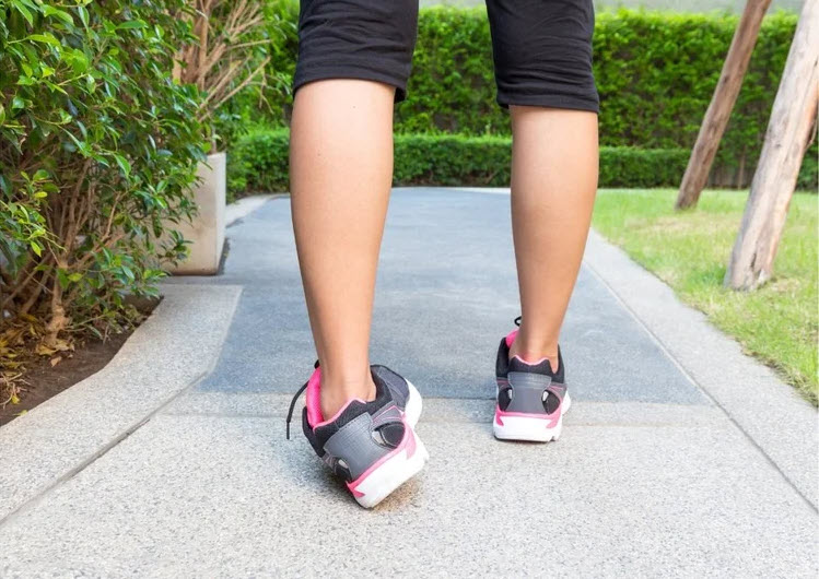
What is an ankle sprain?
An ankle sprain is caused by sudden inversion or eversion, usually combined with plantar flexion or dorsiflexion of the ankle. The ligaments that hold the ankle together become too stretched and sometimes tear. First time acute ankle sprains can lead to chronic ankle instability, which leads to recurring injuries to the ankle. Ankle sprains can happen in just about every sport, contact or no contact. An ankle sprain varies from a mild to severe injury. One of the most common injuries an athlete experiences, is an ankle sprain.
Types of ankle sprains
Inversion ankle sprains are the most common to see. An inversion ankle sprain is caused when the ankle is inverted, plantar flexed and internally rotated position. This type of ankle sprain causes injury to the lateral ligaments of the ankle. Eversion ankle sprains are a lot less common because of the strength of the medial ligaments, specifically the deltoid ligament. An eversion ankle sprain happens when the foot is forced into over pronation. A syndesmotic ankle sprain, also known as high ankle sprain, is an isolated injury to the tibiofemoral joint. This type of sprain occurs when the ankle has increased external rotation or forced dorsiflexion, it normally comes along with a sprain to the medial and lateral complexes.
Ankle sprains are graded based on severity.
- Grade 1: most common, overstretched ligaments. mild pain and disability occur, some swelling may be present and weight bearing is minimally impaired.
- Grade 2: some tearing of the ligaments. usually a pop or snap sound is heard, weight bearing is difficult, moderate pain, swelling and bruising are visible.
- Grade 3: complete rupture of the ligaments. pain is severe, weight bearing is not possible, large amounts of swelling and bruising are visible.
Treatment and prevention
An acute ankle sprain can lead to chronic ankle instability. Ankle instability can lead to more injuries which result in longer rehab time for the athlete and increased missed playing time. Ankle injuries can be a result of poor foot mechanics. When the position of the ankle and foot are not in neutral the athlete is more susceptible to an injury when those ligaments are put in a stressed position. Adding custom orthotics to an athlete’s sneakers and cleats rearrange the foot and ankle anatomically into a neutral position. Now when an athlete is playing, the ligaments aren’t overworking to control the excess movement of the foot and ankle because it is in the correct position. This small change significantly decreases the chance of an ankle sprain.
If you have further questions, or would like to set up a time for an examination, visit us at www.drleecohen.com or call our Marlton, N.J. or Ridley Park, PA. location at 610-522-9200.

